
Treponema Pallidum (Syphilis)
Authors: Lori E. Fantry, M.D., M.P.H., Edmund C. Tramont, M.D., F.A.C.P.Syphilis has been known to afflict mankind at least since the discovery of the “New World” by Christopher Columbus. Before the advent of antibiotics, it was one of the most common infections, afflicting up to 10% of the adult populations in the Western World. Although it was one of the first infections to be treated successfully with antibiotics (91), debate remains with regards to what constitutes optimal treatment. The fundamental reason for this controversy is the inability to culture Treponema pallidum in vitro on routine culture media or in tissue culture. The only means of laboratory culture remains in laboratory animals. This requirement has made it extremely difficult to correlate clinical signs and symptoms with the presence or absence of replicating spirochetes or perform simple in vitro antimicrobial susceptibility testing which in turn has forced clinicians to rely on imperfect tests to diagnose syphilis and gage the effectiveness of therapy.
MICROBIOLOGY
Syphilis is caused by a thin, tightly coiled spirochete, Treponema pallidum subspecies pallidum (92). It is microaerophilic and cannot grow on standard culture media. It is a member of the family Spirochaeticea and is related to other spirochete genera which have the capacity to infect man, namely Borrelia and Leptospira. Other pathogenic treponemes for man include T. pallidum subspecies pertenue, the causative agent of yaws; T. carateum, the causative agent of pinta; and T. pallidum subspecies endemicum, the agent associated with non-venereal or endemic syphilis.The entire genome of T. pallidum was completely sequenced in 1998 (30). It is a small genome of only 1,138,006 base pairs and 1041 predicted coding sequences (open reading frames). Because of this, it lacks many pathways including the tricarboxylic acid cycle, components of oxidative phosphorylation, and most biosynthetic pathways and relies on the host to perform necessary functions (69).
EPIDEMIOLOGY
Although syphilis can be spread by passage through the birth placenta (congenital syphilis), by kissing or other close contact with an active lesion, transfusion of fresh human blood, or by direct inoculation, the vast majority of cases are transmitted by sexual contact (92). Persons are most infectious early in disease when a chancre, mucous patch, or condyloma latum is present and by 4 years after acquiring the disease, an immunocompetent person is essentially non-infectious. Reinfection is possible and, in fact, not uncommon (28).The disease is worldwide in distribution and the World Health Organization (WHO) estimates that there were 12 million new cases of syphilis in 1999 (96). It occurs primarily in persons between the ages of 15 and 40 years but unborn children and 1 million infants are born each year with congenital syphilis.
In the United States, prior to the advent of penicillin treatment, up to 10% of persons living in urban areas were infected (91). The incidence plummeted to very low levels by the mid-1950s and kept decreasing until the year 2000 when the incidence reached an all time low (11). In fact, in October of 1999, Surgeon General David Satcher announced the Center for Disease Control and Prevention’s (CDC) National Plan to Eliminate Syphilis (8).
However, since 2000 there has been an annual increase in the rate of primary and secondary syphilis in the United States (9). This increase was initially fueled by increases among men who have sex with men and, in fact, there were no increases among women but since 2004 the incidence of syphilis has also increased among women. The seroprevalence is greatest in the South, urban areas, among African Americans, and in persons with lower income and less education (31).
CLINICAL MANIFESTATIONS
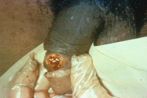
The clinical disease manifestations have been well characterized for over 100 years and are traditionally divided into five stages: incubating, primary, secondary, latent (early latent and late latent), and late or tertiary syphilis (neurosyphilis, cardiovascular syphilis and gummatous syphilis) (92).Primary syphilis is the stage of infection which occurs 3 to 90 days (a median of 3 weeks) after infection. It is most commonly characterized by a single, painless chancre or ulcer that develops at the site of inoculation (see Figure 1) (92). The chancre typically has a smooth base with raised and firm borders. However, in some persons, it does not develop at all; in other persons, it is so small that it may go completely unnoticed; and in others, especially those with human immunodeficiency virus (HIV) infection, multiple ulcers may develop (78,79). The untreated lesion or lesions usually heal spontaneously in 2 to 8 weeks (range 1 to 12 weeks).
Secondary syphilis, the result of the interaction between a large spirochete load and the immune response, is the stage of infection in which there is widespread dissemination to various parts of the body. It becomes evident a mean of 6 weeks (range 2 to 16 weeks) after inoculation. In 90% of cases, there is a rash which most commonly is widely disseminated, maculopapular and involves the palms and soles but other dermatological manifestations are also common (see Figure 2), In addition, over 50% of cases have fever, malaise, anorexia, weight loss, pharyngitis, laryngitis, and/or arthralgias . Other clinical manifestations include lesions in the mouth and oral cavity, lymphadenopathy, condyloma latum, glomerulonephritis, nephritic syndrome, hepatitis, arthritis, osteitis, and periosteitis.
Latent syphilis is the period of months to years in which there are no outward clinical manifestations of disease despite viable organisms. Clinical relapses can occur during the first year of the latent stage (referred to as the early latent phase) and is felt to be the result of waning cellular immunity.
Tertiary or late syphilis occurs in up to 35% of untreated patients ten to twenty-five years after the initial infection. Late syphilis can be categorized into neurosyphilis, cardiovascular syphilis, and granulomatous syphilis. All of these types of late syphilis are uncommon in the antibiotic era except neurosyphilis, reflecting the generally poor penetration of antibiotics into the CNS.
LABORATORY DIAGNOSIS
Because of our inability to culture T. pallidum using standard laboratory methods, a variety of tests have been developed to try to overcome this shortcoming (Table 1).Dark Field Microscopy
Dark field microscopy is still the most sensitive, direct, and quickest method for diagnosing syphilis (92). However, it requires a special dark field equipped microscope and skilled technicians that are not usually available at most medical facilities today, including many large hospitals in the United States. Specimens for microscopic examination are best obtained from serous transudate of moist lesions such as such as a primary chancre, condyloma latum (wart like lesions, sometimes extensive), or mucous patches (shallow ulcers on mucous membranes, non-painful unless secondarily infected). However, they also can be collected from dry skin or lymph nodes by non-bactericidal saline aspiration. When obtaining a sample, the surface should be cleaned with non-bactericidal saline and gently abraded with dry gauze, just enough so that a few scattered red blood cells are seen on the slide. The cleaning should be performed without soap, a topical antiseptic or bactericidal saline because dead and non-motile organisms are difficult to visualize. The specimen should be placed on a glass slide with a cover slip placed on top. When using serous exudates, a drop of non-bactericidal saline may be added if the serous preparation is too thick. Under dark field microscopy, T. pallidum will appear as corkscrew shape in spiraling motion with a 90o undulation about its midpoint. At least three specimens should be examined before deciding that a lesion is non-syphilitic.Nontreponemal Reaginic Antibody Tests
Syphilis reaginic antibodies are IgG and IgM antibodies directed against a lipoidal antigen resulting from the interaction of host tissues with T. pallidum or from T. pallidum itself (92). The earliest cardiolipin antigens used to measure reagenic antibody were crude extracts from beef livers or hearts and false-positive tests were common. However, today’s preparation, the cardiolipin-cholesterol-lecithin, is much purer and hence there are less false-positive reactions. The nontreponemal test first developed was the Venereal Disease Research Laboratory (VDRL) slide test (91). In this test, serum is heated to 56o C and tested to seer if it can flocculate a suspension of a cardiolipin-cholesterol-lecithin antigen. Except for its use in diagnosing neurosyphilis, it has now been largely replaced by the modifications such as the rapid plasma reagin (RPR) card test, automated regain test (ART), and the toluidine red unheated test (TRUST). In addition, there is also now a modified RPR test that can be done without requiring a laboratory at the point of care (93).Nontreponemal tests become positive shortly after initial infection, peak during the secondary or early latent stage, and then decline with time (36) (Table 2). In primary syphilis, an antibody response may not yet have been generated so nontreponemal tests may be negative. In secondary syphilis, virtually 100% of infected persons have positive serological tests for syphilis but in some patients the titers are so high that the test is reported as negative due to the prozone phenomenon (92). If the clinical index of suspicion is high, appropriate serum dilutions should be performed. In latent and late syphilis, titers decline, usually to <1:4, and actually become negative in 25% of untreated persons.
On the other hand, whenever there is a strong immunologic stimulus (e.g., acute bacterial or viral infection, vaccination, HIV infection), a “false” positive occurs (48, 92). In addition, persons who use injection drugs, have autoimmune or connective tissue diseases (especially systemic lupus erythematosus), or hypergammaglobulinemic states may have “false” positive results. These persons often also have blood tests positive for other factors frequently associated with autoimmune disease such as antinuclear, antithyroid, or antimitochrondrial antibodies; rheumatoid factor; and cryoglobulins. A negative specific treponemal test will confirm that the test is a false positive and that syphilis can be excluded.
Fluorescent Treponemal Antibody-Absorption (FTA-abs)
The FTA-abs uses. T. pallidum harvested from rabbit testes as the antigen in a standard indirect immunofluorescent antibody test. The first step is to remove so called “natural” cross-reacting antibody that may have been raised against saprophytic treponemes of the oral cavity or genital tract by absorbing the patient’s serum with nonpathogenic treponemal antigen (referred to as “sorbent”). The next step is to place the patient’s “absorbed” serum on a slide which contains that has fixed T. pallidum as the antigen. If specific antibody to T. pallidum is in the patient’s serum, then it is detected when fluorescein-labeled antihuman gamma globulin is added to the slide and examined under a fluorescence microscope. Its interpretation can be quite subjective. This test is used to confirm or refute a positive nontreponemal test. If there is a high index of suspicion, it also is used to make a diagnosis of syphilis even when a nontreponemal test is negative.T. pallidum Haemagglutination Assay (TPHA) and Microhemagglutination Assay for Antibodies to T. pallidum (MHATP)
The TPHA also measures specific treponemal antibody. It is easier to perform than the FTA-abs and is as specific but not as sensitive, especially in early disease. The MHATP test is similar to the TPHA test except it uses a microtiter plate. “Sorbent” is always used to increase its specificity.Enzyme-linked Immunosorbent Assay (ELISA)
The syphilis ELISAs are automated tests that use a technique called a qualitative sandwich immunoassay to detect T. pallidum specific antibodies. An ELISA may detect only IgG or IgM but most assays are polyvalent. As with all ELISAs, an enzyme is conjugated with anti-human antibodies and only those wells that contain T. pallidum specific antibodies conjugated to the enzyme will exhibit a color change. The sensitivity and specificity of ELISAs are similar to TPHAs and FTA-Abs (7). When compared with the sensitivities of the RPR test and the MHA-TP, ELISA is more sensitive in all stages of syphilis except in secondary syphilis when all tests show 100% sensitivity (7).Immunochromatographic Strip (ICS)
The ICS test is “lateral flow” test in which antibodies in a specimen are detected by becoming bound to antigens, marked with dye, on a cellulose strip. It requires no special training to read, laboratory equipment to run, or refrigeration of reagents or samples. It is ideal for health care facilities in developing countries which lack these resources. In addition, it is takes less than 30 minutes so that treatment, if needed, can be given on the day of diagnosis. A recent study found that tests of this type were 84.5-97.7% sensitive and 84.5-98% specific (38). Line immunoassay (LIA): The LIA uses recombinant and synthetic polypeptide antigens derived from T. pallidum proteins to determine if a clinical specimen has treponemal antibodies (43). In a recent study, the sensitivity and specificity of LIA were 100 percent and 99.3 percent, respectively. Like the ICS test, it is inexpensive, rapid, and requires no special laboratory equipment or highly trained personnel so it is well suited for use in developing countries.Immunofluorescent and Immunoperoxidase Antibody Staining: Specific immunofluorescent or immunoperoxida1e antibody staining can be used to visualize organisms, including nonviable spirochetes, from mucocutaneous lesions, lymph nodes, or dry skin (14, 92). In addition, it can be used for examining non-frozen biopsy material (91).
Polymerase Chain Reaction (PCR)
PCR is available through the CDC but as yet is not available for routine clinical practice in the United States (33,46,52,72). It is most commonly used to diagnose congenital syphilis.Testing Algorithms for Syphilis in Adults except Neurosyphilis
The tradition algorithm for diagnosing syphilis is to screen with a nontreponemal test and then if the nontreponemal test is reactive, then obtain a FTA-abs, TPHA, or MHATP. If both tests are reactive, then a person is considered to have present or past syphilis infection. Treatment decisions are based on past history and nontreponemal titers.Recently, health care facilities have begun using treponemal ELISA for screening because these tests are even cheaper than nontreponemal tests if done in large volumes. Confirmation of current disease that requires treatment is then done with a nontreponemal test (12).
As with the traditional method, when a person is reactive to both the treponemal EIA test and RPR, then a person is considered to have past or current syphilis. When a person is reactive to the treponemal test but nonreactive to the RPR test, persons with a history of previous treatment will require no further management. Those with no prior history of treatment should have a different treponemal test performed such as an FTA-abs. If the second test is also nonreactive, then the clinician needs to decide whether or not a third treponemal test is indicated.
Testing Algorithms for Congenital Syphilis
The diagnosis of congenital syphilis is best made by testing the mother at the time of birth since infant serum titers, even when the infant is infected, may be non-reactive, especially if the mother has low titers or the mother was infected late in pregnancy (10, 92). If the mother has reactive syphilis serology, then the infant’s serum should be evaluated with a RPR or VDRL. Infants should also have a physical exam and dark field microscopy or direct fluorescent staining of any suspicious lesions and radiological and ultrasound studies (10). The placenta or umbilical cord should also be examined using specific fluorescent antitreponemal antibody staining. IgM-specific antibodies [ELISA, reverse enzyme-linked immunospot (Relispot), FTA-abs, or immunoblotting/Western blot] and PCR are also recommended to make the diagnosis (10,59,60,86,92). Any positive tests requires treatment except if the infant’s nontreponemal serology is the same or less than fourfold that of the mother, the mother was known to have been adequately treated with penicillin during pregnancy, and all other tests are negative.Testing Algorithms for Neurosyphilis
A lumbar puncture with the CSF sent for VDRL, cytology, and protein is the most commonly used method for making the diagnosis (10). In the appropriate clinical setting, a reactive CSF-VDRL is considered diagnostic of syphilis while a negative test does not rule out disease, e.g., it is specific but not very sensitive test (92). If the CSF-VDRL is negative, a finding of more than five mononuclear cells per cubic millimeter, a protein value of 46 mg/dL or greater, or a glucose of 45 mg/dL or less all are suggestive of neurosyphilis (18,54). However, the limitations of these tests have been well known for over 80 years and were most recently dramatically illustrated in a 1988 study (54). Four of twelve patients (33%), in whom T. pallidum was cultured from the CSF using rabbits had normal cellular and protein studies, as well as a non-reactive CSF-VDRL (RPR), attesting to the low sensitivity for all of these tests, even in combination. In addition, there were a significant number of patients in whom T. pallidum was not isolated from the CSF despite CSF abnormalities including four patients with reactive VDRL’s. A serum VDRL (RPR) titer greater than 1:32 most closely correlates with a positive T. pallidum isolation in rabbits (culture) and this was found most commonly in secondary syphilis. Other methods with greater sensitivity and specificity than the CSF-VDRL are the intrathecal T. pallidum antibody (ITPA) and TPHA indices but these are seldom used in clinical practice (92). A FTA-abs test is usually not performed on CSF because a positive test may represent passive transfer of antibody from serum to the CSF and not active CNS disease (92). However, it is highly sensitive and thus a negative test can be helpful in ruling out neurosyphilis, especially in HIV infected patients who often have white blood cells in the CSF.. Marra and co-workers found it, along with a determination of a percent CSF cells that were B lymphocytes, to be useful in excluding or establishing a diagnosis of neurosyphilis (58). Clinical judgment must be the deciding factor or treat for neurosyphilis!PATHOGENESIS
Infection begins when T. pallidum penetrates the host, usually through intact or abraded mucous membranes (92). Although the exact infectious dose in humans is not known, in rabbits, an infectious inoculum can be as few as four spirochetes. The incubation period is directly proportional to the size of the inoculum since clinical lesions do not appear until a concentration of approximately 107 organisms per milligram of tissue in rabbits is reached (21). Potential virulence factors which account for T. pallidum’s ability to cause disease include several hemolysins, a membrane protein that allows for permeability of nutrients but inaccessible to antibody, and ligands that allow cytoadhesion (30,37, 73). As with most other infections, it is the ensuing inflammation that is responsible for most of the disease pathology (92).In early disease, spirochetes can be found in the chancre, the usual first manifestation of syphilis. Invasion into the bloodstream and lymphatics occurs within hours to days of penetration of T. pallidum as evidenced by the fact that patients who received blood transfusions from syphilitic donors in the seronegative incubation period have become infected (92). All organs of the body can be invaded but the skin, lymph nodes, and the central nervous system (CNS) are the sites most often invaded. In the skin, T. pallidum is found in the dermal-epidermal junction zone or throughout the dermis (22). Up to 40% of patients in primary and secondary syphilis have evidence of CNS invasion, including the eye, as evidenced by either abnormal laboratory tests or direct culture of the treponemes in laboratory animals. Late stage disease is most often manifested by invasion of the vasa vasorum of the aorta and/or the arteries of the CNS by T. pallidum (92). In addition, gummas, granulomatous lesions with coagulated or amorphous centers, form most commonly in the skin, liver, bones, and spleen.
Both humoral and cell mediated immune responses are mounted against T. pallidum. At all stages of infection, there are local cellular infiltrates consisting of lymphocytes, macrophages, and plasma cells at the sites of disease (73). In primary syphilis, CD4+ T cells and macrophages are the predominate cell type while in secondary syphilis CD8+ cells predominate. In both primary and secondary syphilis, there is increased expression of Th1 cytokines IL-2 and IFN-gamma. Cell mediated and humoral immune responses peak in secondary syphilis.
Spirochetes may remain alive and continue to replicate in immunologically sequestered sites in the body even with a brisk initial immune response and no outward clinical manifestations of disease in at least 1 of 4 persons who are not treated. In late latent syphilis, after many years of latency, treponemes begin to multiple and Th1 lymphocytes produce high levels of nitric oxide and IL-12 instead of IL-2 and IFN (55). This immunological escape by T. pallidum is thought to be due to three main characteristics of T. pallidum. First of all, it has a very slow dividing time of 30-33 hours. Secondly, it has a scarcity of protruding antigenic proteins membrane proteins which make an antibody response more difficult. Thirdly, it has the ability to vary its antigens by gene conversion which may result in a subpopulation of T. pallidum to immunological defenses such as macrophage phagocytosis (73, 92).
SUSCEPTIBILITY IN VITRO AND IN VIVO
Single Drug
Early susceptibility testing of various antibiotics against T. pallidum in experimental rabbit models established the principles of anti-treponemal syphilis therapy. Using the rabbit model, Eagle and co-workers in the 1940’s found that established later stage disease required a longer course of therapy but cure could be accomplished with relatively low dose therapy (21) given over a prolonged period of time. These early studies have served as the basic tenants in our approach to syphilis therapy.Penicillin, the first antibiotic developed, was the first known effective antibiotic for T. pallidum and remains the treatment of choice today (10, 24, 74). The maximally treponemicidal serum concentration of penicillin is 0.36 µg/ml which can kill the organism in 6 to 9 hours (21). However, a concentration as low as 0.005 µg/ml can clear T. pallidum from chancres if it is maintained for 48 to 96 hours (21). In the rabbit model, viable treponemes may persist in the lymph nodes even after early lesions have been cleared. T. pallidum can regenerate if the serum penicillin concentration falls to sub-inhibitory levels for 18-24 hours (42).
Resistance to penicillin has not yet been found among clinical isolates (92) but there is the potential for the acquisition of extrachromosomally mediated antibiotic resistance since at least one strain has been shown to contain plasmid DNA (68).
T. pallidum is also susceptible to virtually all other β-lactam antibiotics. Based on studies done with the time honored in vivo rabbit model, amoxicillin (63), ceftriaxone (51), ceftizoxime (50), cefmetazole (2) and cefetamet (24) have been shown to be curative. Azithromycin, which has the advantage of once daily dosing, also has activity against T. pallidum (53,88).
Macrolide antibiotics are also able to inhibit T. pallidum but not as efficiently as the β-lactam antibiotics and unlike penicillin, clinical resistance to macrolides have been described (42). Thus, they are relegated to second line antibiotic treatment status. For example, when erythromycin was directly compared to penicillin G for the treatment of rabbit skin syphilomas, a single injection of penicillin decreased the motile T. pallidum count by more than 250-fold, whereas single doses of erythromycin did not decrease the count significantly (5). Only after multiple injections of erythromycin was the number of motile treponemes decreased in amounts similar to that of penicillin. Chloramphenicol, an antibiotic no longer available in the USA, also has activity against T. pallidum but has failed to eradicate infection in the rabbit model (42). Never the less, because it concentrates in the CNS, it has utility in treating neurosyphilis. Spectinomycin (74), the quinolone class of antibiotics (90), and clindamycin (5) have negligible activity against T. pallidum and are contraindicated.
Combination Drugs
Because of the exquisite success of single drug therapy, combination therapy for syphilis is not required and has not been studied.ANTIMICROBIAL THERAPY
The following points are important to note before a discussion of antimicrobial therapy for syphilis. First, as in the case of many infections, a natural immune response can clear T. pallidum independent of antibiotic usage. On the other hand, in states of immune impairment, especially with cell-mediated deficits such as occurs in the Acquired Immunodeficiency Syndrome (AIDS), even the most effective antibiotics may fail to eradicate T. pallidum (56). Hence, antibiotics augment clearance and subsequent cure but do not guarantee it. Furthermore, the pharmacology of the antibiotic preparation must be considered, specifically whether it reliably reaches the CNS in adequate amounts to inhibit T. pallidum because the CNS is invaded in up to 40% of patients (54). Unfortunately, many of the current therapies recommended for syphilis do not sufficiently penetrate into the CNS to best assure a cure of incubating neurosyphilis (19, 20, 61).Penicillin is the best studied and still remains the recommended therapy for syphilis (91). However, after nearly 60 years, there still remains much debate as to what dosing schedules and which preparations are appropriate for the various stages and types of persons infected with T. pallidum (76). The original studies of penicillin treatment were performed before the establishment of the standard double-blinded placebo or comparison controlled randomized clinical trials to evaluate the efficacy of therapy. In other words, there have been no rigorous studies of penicillin or any other antibiotics to evaluate dose, duration, and preparation of antibiotic that would achieve cure with minimal toxicity and cost. Hence, all recommendations of treatment for syphilis must be regarded with a degree of skepticism.
Because therapy should be tailored according to the benefit that a person will receive from a particular therapy balanced with the inconvenience and cost of alternative forms of therapy, the following discussion will focus on therapeutic preferences rather than rigorous recommendations.
2006 CDC Recommendations
The CDC periodically reviews the recommended treatments for syphilis (Table 3) (10). The CDC treatment guidelines are based on the premise that different stages or forms of the disease should be treated with different preparations and doses of penicillin. Following the principles of public health or population based guidelines, their goal is to provide therapeutic recommendations that will cure the greatest number of patients with minimal expense, easy administration, and high compliance (42). In contrast, treatment of the individual patient rests with the treating clinician who must weigh the risk of failing to cure a curable disease.The CDC recommends that non-pregnant women and men with early syphilis, which includes primary syphilis (the stage that occurs immediately after infection characterized by an ulcer or chancre); secondary syphilis (the later stage corresponding to spirochete dissemination and characterized by skin rash); and early latent syphilis (the stage up to one year after initial infection when positive serology’s are the only indication of infection), should be treated intramuscularly with one injection of 2.4 million units of benzathine penicillin G (10). This is based primarily on animal data showing that the minimal inhibitory concentration that cures primary lesions is easily achieved with this dose of benzathine penicillin (42). However, this does not take into consideration the fact that in 40% of cases of early syphilis there is CNS invasion which can result in the development of late neurosyphilis (54) and that this dose of benzathine penicillin does not reliably penetrate the CNS (19, 20, 61, 75, 98). Furthermore, a failure rate of up to 5% was suggested in the early clinical trials.
The CDC also recommends that non-pregnant women and men with late latent syphilis (syphilis of more than 1-year in duration), latent syphilis of unknown duration, and tertiary syphilis except neurosyphilis) also receive benzathine penicillin (Table 2) but at an increased total dose of 7.2 million units given as 2.4 million units weekly for three weeks (10) to account for the possible longer dividing time of the spirochete at this stage of disease (42). However, as noted above, CNS concentrations of benzathine penicillin are not reliably achieved (54).
Acknowledging the well-recognized fact the neurosyphilis is the most difficult manifestation to treat, the CDC recommends that persons with neurosyphilis be treated with 3 - 4 million units of aqueous crystalline penicillin G given intravenously every 4 hours (18-24 million units daily) for 10-14 days. However, who should be included in this classification of “neurosyphilis” is vague and ill defined. Patients with a reactive cerebrospinal fluid VDRL or RPR test are the easiest to classify and are accepted as definitely having neurosyphilis. But most patients have either no symptoms or ill-defined symptoms and most have nonspecific CSF abnormalities. Furthermore, despite the fact thatT. pallidum invades the CNS in 40% of patients (54), a lumbar puncture is seldom performed unless the signs and symptoms of acute meningitis are manifested as part of the signs and symptoms of secondary syphilis. In fact, the CDC discourages performing a lumbar puncture in early syphilis unless the patient has optic, auditory, cranial nerve, or meningial symptoms (10). The rationale is that although early invasion of the CNS by T. pallidum is common, it does not predict treatment failure with benzathine penicillin and that the lumbar puncture often yields little additive information.
In summary, the CNS is invaded soon after infection in up to 40% of patients and some will go on to develop late neurosyphilis 10-25 years later. Unfortunately, the diagnosis is difficult to establish, and benzathine penicillin does not reliably reach the CNS in the majority of cases, setting the stage for well-documented treatment failures. The clinician must weigh the risk of CNS invasion in each individual patient.
Treatment Failures
Although benzathine penicillin is effective therapy for the vast majority of persons with syphilis, there are multiple reports of treatment failures, especially in patients co-infected with Human Immunodeficiency Virus (HIV). It has long been established that benzathine or low dose aqueous or procaine penicillin is not 100% successful in preventing neurosyphilis in non-HIV infected persons, so it not surprising that these immune dysfunctional patients would be particularly vulnerable to this complication, especially when one considers that re-treatment is necessary in at least 5% of immunocompetent patients with primary or secondary syphilis treated with low dose aqueous benzathine procaine penicillin (6, 76, 83, 84). Although this failure can be at times attributed to re-infection (25, 26), at least half of those cases are convincingly due to failure of initial therapy (83).The most likely reason for treatment failures is that at least 40% of patients with early syphilis are infected with T. pallidum in the CNS (53), a site that is poorly penetrated by benzathine penicillin (19, 20, 27, 47, 54, 61, 65, 75). In most cases of syphilis, the CNS infection and poor penetration of penicillin is inconsequential since an immunocompetent host is usually capable of clearing the infection before any neurological damage occurs.
Since the most reliable form of treatment is one that is reaches treponemicidal levels in the CNS, the most reliable preferred therapy for any form of syphilis in any type of patient is 2 - 4 million units of aqueous crystalline penicillin G given intravenously (iv) every 4 hours (12-24 million units daily) for 10-14 days (Table 4) since between 5 and 24 million units of intravenous penicillin G consistently achieves levels in the CSF that are treponemicidal (61,75). Hence, it would follow that treatment failures would be rare when compared to regimens that failed to reliably penetrate the CNS (64).
Oral Regimens
Because inpatient hospital treatment is not always practical from a reimbursement or lifestyle perspective, other treatment regimens have been developed. The oral regimen most thoroughly studied is the semi-synthetic penicillin, amoxicillin, given 3.0 gm orally twice a day with probenecid 500 mg orally for 10-14 days (See Table 3). Probenecid inhibits renal excretion of natural and semi- synthetic penicillins. As previously noted, amoxicillin is treponemicidal in the rabbit model , and when 2 gms of amoxicillin plus 500mg of probenecid is administered to fasting adults, the MIC for T. pallidum can be exceeded for at least eight hours (23, 63). Clinical trials, albeit small, have confirmed the utility of this regimen (23,70,77).Both of these regimens can be used in adults at any stage of syphilis. However, in neurosyphilis, even these regimens may fail and re-treatment may be required (95), especially in HIV infected patients (65). Hence, diligence of follow-up must be the order of the day.
Unfortunately, reversal of late neurological deficits due to syphilis is rarely obtained (42).
Alternative Therapy
2.4 million units of aqueous procaine penicillin G have been given intramuscularly daily with probenecid 500mg orally 4 times per day for 10-14 days. Treponemicidal levels of penicillin have not been achieved as reliably with procaine penicillin but supplementation with probenecid will achieve treponemicidal levels (20). Furthermore, patient compliance is problematic because of the pain associated with the injections. Since this therapy avoids the need for frequent dosing and thus hospitalization, it is recommended as an alternative in compliant patients in whom the local discomfort is tolerable.The first- and third- generation cephalosporin antibiotics, in particular ceftriaxone, are also effective in treating syphilis. Ceftriaxone has been recommended for treatment of early disease and neurosyphilis (Table 3) (10). This drug is particularly attractive since serum and CSF levels exceeding those needed to kill T. pallidum are easily obtained with standard dosing (51). It has been used clinically to successfully to treat incubating syphilis (42), primary syphilis (42,62,82), secondary syphilis (42,82), latent syphilis (18), and neurosyphilis (57, 74). However, there have been reports of treatment failures in HIV infected patients, similar to those found with benzathine penicillin (18).
Doxycycline (100 mg twice daily for 21 days) is also an effective alternative for treatment of the non-HIV infected adult with syphilis. The enthusiasm for this regimen is also restrained because it is bacteriostatic in vitro and not bacteriocidal (92). Furthermore, tetracycline and doxycycline are contraindicated in pregnant women and children under the age of 8 because of potential damage to developing bones and teeth (67). Since tetracycline and doxycycline have similar spectrums of antibacterial activity, most data in support of doxycycline has been extrapolated from previous trials with tetracycline, a drug that one is less inclined to recommend because of its poor CSF penetration. Patients treated with 24-32 grams of tetracycline for 10 -12 days have had similar serological and symptomatic response to therapy when compared to penicillin (25,26,83). One study reported a failure rate at 12 months of below 4%, which was similar to that obtained with benzathine penicillin therapy (82). However, one small prospective observational study showed that doxycycline (100 mg bid for 28 days) resulted in 100, 90, 68, and 90% success rates in treating primary, early, late, and congenital syphilis, respectively (71).
Other regimens that are less desirable because of poor CSF penetration include benzathine penicillin, as previously discussed, and the macrolide antibiotics. Erythromycin had a failure rate of 14% after a follow up of 18 months (82). Another macrolide, azithromycin, has been shown in a recent meta-analysis to be more effective than benzathine penicillin G for treating early syphilis (1). However, in addition to its probable inadequate penetration into the CSF (85), it has been associated with resistance and treatment failures (49) and therefore it can only be recommended when there are no other alternatives. Because of excellent CNS penetration, chloramphenicol, when available, (2 gm daily for 30 days) is another alternative that can be used for treatment of neurosyphilis. In every instance, prolonged treatment will result in a cure since, with the exception of erythromycin, antibiotic resistance has not yet evolved.
Special Situations
Congenital Syphilis
For newborns, the treatment is 50,000 units/kg of aqueous crystalline penicillin G intravenously every 12 hours for a total of 100,000-150,000 units/kg/day for the first 7 days of life and then every 8 hours for a total of 10 days (10). The alternative regimen is 50,000 units/kg of procaine penicillin intramuscularly daily for 10 days. If treatment is interrupted for more than 24 hours, then the full course of therapy is restarted. Children diagnosed beyond the newborn period with congenital syphilis should also be treated presumptively for CNS involvement with therapy as described above except that aqueous crystalline penicillin needs to be administered every 4-6 hours instead of every 8 or 12 hours (86).Optic Syphilis
Syphilitic involvement of the eye, a relatively common occurrence in HIV co-infected persons, should be treated with penicillin G 12 to 24 million units per day for 10 to 14 days regardless of CSF findings (15). Penicillin, however, regardless of dose or preparation, does not easily achieve treponemicidal levels in anterior chamber of the eye and hence, requires high doses and prolonged treatment (14 days) to improve the chances of adequate aqueous fluid levels (92). An illustrative case is that of a newborn with congenital syphilis receiving penicillin G potassium 50,000 units/kg body weight given intramuscularly at 12 hour intervals for 17 days after birth and T. pallidum were found in both the aqueous humor and ground eye tissue at autopsy (35). Hence, it may also be prudent to consider chloramphenicol or cephalothin, antibiotics that do achieve adequate levels in the anterior chamber of the eye, as possible first line agents (92).Otosyphilis
As is the case of penetration into the anterior chamber of the eye, penicillin does not easily penetrate the labyrinth of the inner ear (92). Hence, a neurosyphilis regimen is warranted. Patients are usually treated with a prolonged course of penicillin for six weeks to three months with the addition of steroids (13, 17, 99) Oral prednisone 30-60 mg daily or every other day for at least 7 to 8 days should be given as tolerated by the patient (99). The utility of the steroids has never been proven but has become the standard of care to guard against the sudden worsening of hearing loss attendant to an acute reaction to released antigens form lysing organisms (Jarish-Herxheimer Reaction).HIV Infected Patients
More florid clinical manifestations, including multiple and more frequent genital ulcers (80) and symptomatic neurosyphilis, occurring early in the course of syphilis suggestive of a high spirochete burden (47), have been well documented in persons co-infected with HIV. In addition, multiple case reports suggest that HIV infected patients are more likely to fail antibiotic therapy than non-HIV infected patients (4, 47, 54, 66). For example, T. pallidum was isolated in three of three HIV infected patients evaluated after single dose benzathine penicillin therapy for early syphilis but could be cultured in the only patient without HIV infection (54). In addition, in a study of 59 patients with neurosyphilis, HIV infected patients were 2.5 and AIDS patients were 3.7 times less likely to normalize CSF-VDRL (58). Multiple retrospective chart reviews have documented treatment failures in up to 20% of HIV infected patients (18, 56), significantly higher than the 5-10% failure rate reported prior to the AIDS era. Furthermore, unlike many treatment failures in immunocompetent hosts, these failures are more likely to be associated with clinical disease, especially neurosyphilis (16, 18, 56).More florid clinical manifestations and the higher frequency of treatment failures are most likely due to immune dysfunction coincident with HIV that results in a significantly higher spirochete load. There is a greater likelihood that spirochetes will remain active in sequestered sites. Hence, in HIV infected patients, it is prudent to use only antibiotics that more reliably penetrate the CNS, such as high dose penicillin or amoxicillin with probenecid, for 10-14 days.
Jarisch-Herxheimer reaction
Treatment for syphilis is not necessarily benign. Along with the discomfort of intramuscular or intravenous therapy and the possibility of a drug reaction, an acute febrile response to therapy is seen in 10-25% of all patients and 70-90% of patients with secondary syphilis (92). It is rarely seen in latent syphilis and occurs in varying amounts in the various forms of tertiary syphilis (81). This reaction, called the Jarisch-Herxheimer reaction, is most commonly associated with penicillin therapy but also has been associated with other antibiotics used to treat syphilis (3). It has been correlated with the release of heat stable “pyrogens” from T. pallidum and other spirochete infections (92) probably resulting in a rapid high release of inflammatory cytokines and chemokines. The clinical course is marked by the abrupt onset of symptoms usually beginning within two hours, occasionally as late as 24 hours, after initiation of therapy (10, 92). Besides fever, clinical signs and symptoms may include chills, headache, nausea, vomiting, myalgias, tachycardia, hyperventilation, mild hypotension, vasodilatation with flushing, and the exacerbation and new onset of skin lesions (81). It lasts 12-24 hours and is generally safe and self-limited ( 92). The exceptions are during the second half of pregnancy when there a risk of premature labor and fetal distress (10) and during cardiovascular, neurosyphilis and otosyphilis when reactions are more severe and can be life threatening (92). Acetaminophen or aspirin can be used for symptomatic relief. In severe cases or those associated with cardiovascular syphilis or neurosyphilis, prednisone 60 mg can be given. When given intravenously, this usually results in a prompt and dramatic decrease in fevers (92).Penicillin Desensitization
Since penicillin is the optimal therapy for syphilis, penicillin should be seriously considered even in patients with penicillin allergies, except in those with a history of Stevens-Johnson syndrome, or alternatives are limited. These situations include syphilis in pregnant women, infants with congenital syphilis and adults with symptomatic neurosyphilis since treatment must be effective as soon as possible to prevent irreversible neurological disease. In addition, in syphilis in pregnancy and congenital syphilis, the toxicities associated with the tetracyclines make this option undesirable not only in terms of possible treatment failures but also in terms of damage to bone and teeth development (67).Allergic skin reactions to penicillin are the principle toxicity associated with this antibiotic, are much less frequent with the pure preparations used today and occurred in at most 4% treatment courses in the past with less pure product (94). In addition, the vast majority of patients who report a history of penicillin allergy will not have an allergic reaction upon re-challenge.
Anaphylactic reactions are extremely rare today. The most reliable method of distinguishing those who will have a future reaction from those who will not is skin testing. Prior to skin testing, patients should be instructed not to take antihistamines, tricyclic antidepressants, and adrenergic drugs within a time interval appropriate for the corresponding half-life of the medication (10). For instance, chlorpheniramine maleate or terfenadine should not have been taken within 24 hours; diphenhydramine HCL or hydroxyzine within 4 days; and astemizole within 3 weeks. Testing is begun by injecting skin test reagents, including a negative diluent and positive histamine controls, in the epidermis on the volar surface of the forearm with a 26 gauge needle without drawing blood (94). If there is no reaction, then the same reagents are injected intradermally and if the diameter of induration is greater than 5 mm after 15 minutes, then the test is positive.
Optimally, skin testing should include diluents with both the major and minor determinants of penicillin (94). If the skin test is negative, then there is no risk of anaphylaxis but 1-3 percent of patients will develop a mild cutaneous reaction. If the skin test is positive, then desensitization should be carried out prior to treatment since there is a 50-70% risk for an acute reaction. Because minor determinants of penicillin are not commercially available in the United States, skin testing is often done with the major determinants only. In this case, if the test is negative then patients should still receive gradually increased doses of penicillin in monitored setting since there still is at least a 3% risk of a reaction.
Although desensitization can be done both orally and intravenously, oral desensitization is probably safer and certainly less expensive and easier (94). It should be performed in an intensive care setting with continuous electrocardiogram monitoring. Doses of penicillin should be gradually increased until the dose required for therapy is achieved. One-third of patients will develop a transient allergic reaction during desensitization but it is usually mild and self-limited. If mild symptoms occur, then the dose of penicillin should be stabilized until there are no signs or symptoms. If it is more sever, such as hypotension, laryngeal edema, or asthma, then the dose should be reduced at least ten-fold. There should be no interruption between desensitization and treatment since there the risk of an allergic reaction increases as the time from desensitization increases.
ENDPOINTS FOR MONITORING THERAPY
Non-HIV Infected Patients
As in any disease, the primary endpoint of therapy is clinical and a carefully taken medical history, physical exam and appropriate laboratory tests will determine if therapy has been successful. In primary syphilis, the patient should be examined for resolution of the chancre; in secondary syphilis, for resolution of the skin rash and any of the other protean disease manifestations that the patient may experience; and in early neurosyphilis for resolution of signs and symptoms of neurological disease. In late neurosyphilis, there is usually no reversal of signs and symptoms that are present prior to therapy but patients should be monitored carefully for further progression of disease. During any stage of syphilis and at any time, if there is persistence or recurrence of clinical disease, the person should be retreated. He or she should also be questioned about the possibility of re-infection to ease the concern about antibiotic resistance.However, since the disease process is clinically silent at many stages and we are unable to grow in routine culture, it is necessary to follow disease activity with surrogate markers of infection. Treponemal antibody tests such as the FTA-abs are not helpful for measuring response to therapy since they are not quantified, usually remain positive for life, and do not correlate with disease activity (78, 87). In contrast, nontreponemal tests such as the RPR and VDRL are quantifiable, revert to negative in the vast majority of patients given effective therapy, and do correlate with disease activity. In fact, without therapy, 25% of patients eventually have non-reactive VDRL tests (36). With therapy, nearly 100% of non-HIV infected persons change from reactive to non-reactive.
Fiumara found that all 196 patients with treated primary syphilis had non-reactive RPR titers at one year (25) and all 204 patients with treated secondary syphilis by two years (26). In addition, he found that only 44% of 275 patients treated for late latent syphilis became seronegative (27). In another series, all patients with primary syphilis had resolution both clinically and serologically but only 55% of patients with secondary syphilis (64). However, some studies have shown that low but positive titer VDRL or RPR tests of 1:16 or less may persist despite no other evidence of treatment failure (the “serofast reaction”) (10,83). This may represent a false positive result or, especially when the titer is >1:4, persistent active infection or reinfection (92). HIV-infected patients are more likely to have a persistently high-titer RPR or VDRL.
Since VDRL titers are often slightly lower than RPR titers and there is significant variation in values between laboratories, either a RPR or VDRL, is best obtained from the same laboratory each time to monitor therapy (10). A fourfold change in titers, equivalent to a two dilution change, is considered a significant change and implies a change in disease activity. For instance, a change from 1:64 to 1:8 is significant while a change from 1:64 to 1:32 is not. The frequency of obtaining serological testing after treatment depends on the stage of disease and characteristics of the patients. Non-HIV infected adults with syphilis should be tested at three, six, and twelve months (92). If syphilis is greater than one year in duration or of unknown duration, then serologic testing should also be checked at 24 months. Pregnant women who are infected should have follow up serology in the third trimester and at delivery (10). If a pregnant woman is at a high risk for re-infection or in an area where the prevalence of syphilis is high, she should be retested every month (10). Infants with congenital syphilis and those who are sero-positive only because of passive transfer of maternal antibodies and are not infected should be checked with serological testing every two to three months (10).
The rate of decline after treatment of nontreponemal titers appears to be related to the stage of disease, height of the initial titers, and a prior history of syphilis (6, 25-28). Brown and co-workers found a fourfold decrease in RPR titers at 6 months and eightfold decrease at 12 months in successfully treated patients with primary and secondary syphilis but there was a slower decline in patients with early latent disease and longer duration of symptoms (6). In contrast, patients with no prior history of syphilis (re-infection) have a more rapid decline in titers (6,25, 26, 29). In addition, patients with macular rashes during the secondary stage revert to negative quicker than those with papular rashes (28). When titers do not decline fourfold by six months in primary or secondary syphilis or patients have a positive RPR/VDRL reaction beyond 12 months in primary syphilis, 24 months in secondary or latent syphilis, or 5 years in late syphilis, then patients should have a CSF examination and be retreated (74).
In adequately treated congenital syphilis, titers should decline by three months of age and should be non-reactive by six months of age (10). Titers, however, may decline slower if treatment is not initiated in the neonatal period and, occasionally, titers are still positive up to one year after birth. If titers are found to be stable or increasing or are still present after one year of age, then the child should be re-evaluated fully, including a CSF examination, and retreated.
Besides following serum nontreponemal test, a lumbar puncture is recommended every six months after treatment for neurosyphilis, including infants with abnormal CSF analyses, until the pleocytosis has resolved or CNS non-treponemal antibodies disappear (92). Pleocytosis usually resolves before CSF protein declines and CSF-VDRL tests may remain reactive for more than one year (47). If the CSF cell count is not decreased by six months or remains abnormal two years after therapy, re-treatment is indicated (8). In addition, in infants if the CSF-VDRL is still positive at six months of age, the infant should be retreated.
HIV Infected Patients
Although HIV infected patients may have similar non-treponemal (VDRL and RPR) response as non-HIV infected persons (32, 45), HIV infected patients are more prone to have a delayed serological response to infection or therapy and greater likelihood of having persistently reactive non-treponemal titers after treatment (95). Furthermore, HIV infected patients are more likely to have high non-treponemal antibody titers in secondary syphilis, e.g. >1:128 dilution (44), an increased rate of false positive non-treponemal antibody titers (35) and are less likely to have a fourfold decrease in titers within 6 months than matched non-HIV infected patients (89, 97). In addition, specific treponemal tests (FTA-ABS, TPHA and MHA-TP) more often become non-reactive in HIV infected patients than in non- HIV infected patients and, as would be expected, this loss of reactivity is associated with lower CD4 lymphocyte counts (34,45).This abnormal response to therapy is most likely due to immune dysfunction related to the increased polyclonal B cell activation found in HIV infection rather than a true change in disease activity, although the resultant loss of effective T-cell function could result in less ability to clear the spirochete (89).
VACCINES
No vaccines are currently available.PREVENTION
The most effective prophylaxis against contracting syphilis is avoidance of sexual contact with persons who harbor the spirochete. Condoms use during sexual intercourse can also be inferred to be protective from studies of HIV infection. Currently, prophylactic drugs are not recommendations in any group of patients with syphilis except those who have been exposed to a person with syphilis and in this population, it is unclear whether medicines prevent infection or treat very early disease, e.g. “incubating” syphilis.We recommend that persons sexually exposed to primary, secondary, or latent syphilis of less than 1- year’s duration within the previous 90 days be treated for syphilis even if they have negative serological tests for syphilis because there is data to suggest that therapy is most successful when initiated early and the treponemal burden is relatively low (35). This is the one instance where treatment with 2.4 million units of benzathine penicillin is acceptable. A possible alternative is a single 1.0-g dose of azithromycin (40).
Transmission occurs only when mucocutaneous lesions are present, which is usually within one year after initial infection (92), so contact with persons with syphilis greater than one year in duration is not an immediate indication for treatment (10). Instead, it is recommended that these persons be followed clinically and serologically for syphilis. Persons with exposure greater than 90 days prior to evaluation need not have immediate treatment, unless they have positive serological test, serological results will be delayed or follow up is uncertain.
Blood transmission is rare today because of the low incidence of disease and blood storage procedures
Post a Comment